
Ultrasound
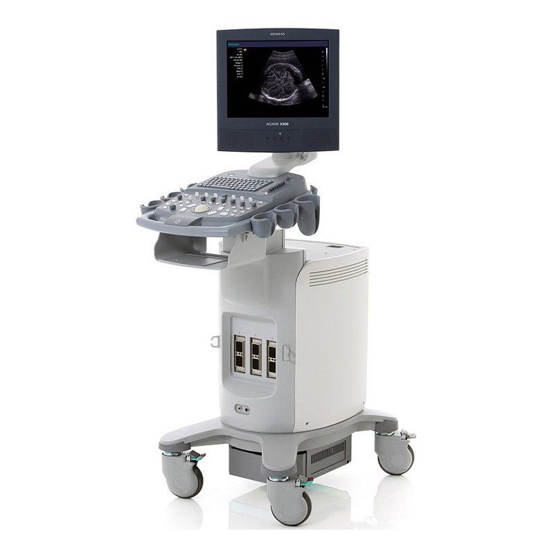
A GE Logiq E9 ultrasound machine has been installed for X-ray examinations.
- Depth of display: 0-36 cm
- Minimum depth of field: 0-2 cm
- Maximum depth of field: 0-36 cm
Benefits
The GE Logiq E9 is a shared ultrasound machine capable of premium gynecological imaging and 4D imaging along with clear imaging for radiology, and vascular applications. GE’s breakthrough technology with the Logiq E9 is XDclear, an imaging technology that uses advanced single-crystal transducers, acoustic amplification and cooling technology to improve image quality, especially at deeper penetration when image quality typically degrades.
Examinations conducted
The upper and lower abdomen include the liver, gall bladder, spleen, pancreas, kidneys, adrenal glands, prostate (for men), bladder and for women the uterus and ovaries.
Upper and lower abdominal ultrasound can provide information on abdominal algae, cholelithiasis, nephrolithiasis, prostatic hypertrophy, uterine lesions, while it can also diagnose early the presence of possible neoplasms. It is an examination of first choice for highlighting alterations without burdening the examinee’s body.
Preparation is required the day before the exam. The examinees must not have consumed fruits, legumes, vegetables, dairy products. They should be fasting for the last 6-8 hours before the examination and also they should have drunk 6 glasses of water 1 hour before the examination and not have urinated, for the best study of the lower abdomen – small pelvis.
The ultrasound of the thyroid gland, as an examination of choice, studies its dimensions as well as the presence or absence of nodules and parenchymal inhomogeneity in it (autoimmune thyroiditis – Hashimoto’s), while it also helps to highlight pathologically enlarged cervical lymph nodes as well as their evaluation.
Thyroid – parathyroid image control after surgery.
The ultrasound of the musculoskeletal system – soft tissues reveals palpable skin formations (skin cysts, lipomas), tendonitis, contusions, hematomas in the soft tissues – ligamentous elements after injuries – injuries. Particularly useful after sports injuries in combination with other imaging methods.
Συμπληρωματική εξέταση συνδυαστικά με την Μαστογραφία που βοηθά στην πρόληψη και την διαφοροδιάγνωση αλλοιώσεων των μαστών.
Διενεργείται ανεξαρτήτως ηλικίας και δεν επιβαρύνει τον οργανισμό των εξεταζομένων.
Carotid and vertebral arteries, arteries and veins of upper and lower extremities, iliac arteries, renal arteries, abdominal aorta.
Early detection of the presence of clots for their immediate treatment. Identification and degree of stenosis of arteries. Aneurysmal distention control and monitoring as well as lower limb swelling.
It depicts in more detail changes of the uterus and ovaries through the vagina. It does not require special preparation.
Emphasis is placed on the endometrium and helps to better highlight and monitor uterine fibroids compared to transabdominal ultrasound.
Emergence of inflammations (epididymitis etc.) varicocele – hydrocele as well as presence of formations in the testicles and scrotum.

This post is also available in el.